


Question
Question asked by Filo student
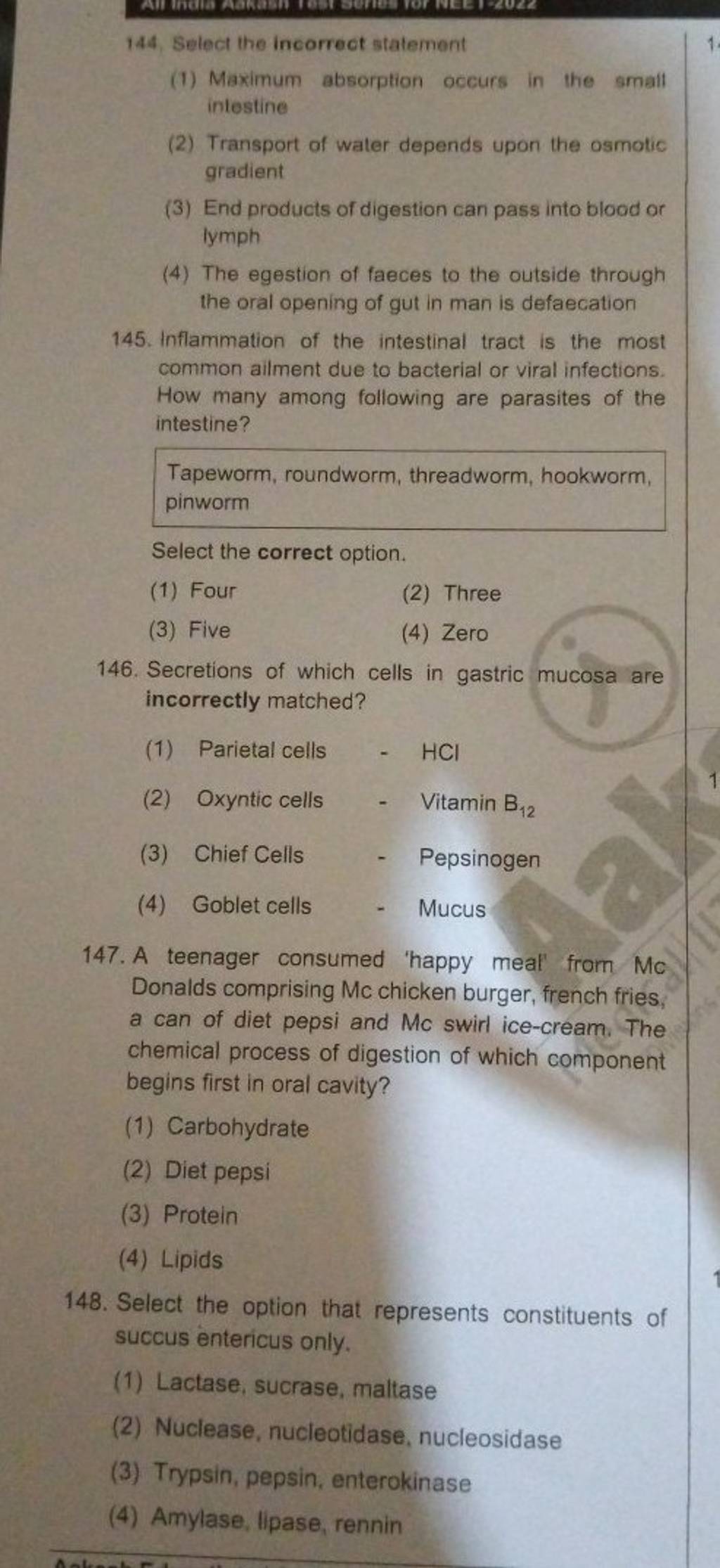
Select the option that represents constituents of succus entericus only.
- Lactase, sucrase, maltase
- Nuclease, nucleotidase, nucleosidase
- Trypsin, pepsin, enterokinase
- Amylase, lipase, rennin
Views: 5,790 students
Found 5 tutors discussing this question
Discuss this question LIVE
15 mins ago
Filo tutor solutions (1)
Learn from their 1-to-1 discussion with Filo tutors.
23 mins
Uploaded on: 12/17/2022
Connect instantly with this tutor
Connect now
Taught by

Mohd Sultan
Total classes on Filo by this tutor - 13,538
Teaches : Biology, Physical Chemistry, Organic Chemistry
Connect instantly with this tutor
Was this solution helpful?
75
Share
Report

One destination to cover all your homework and assignment needs
Learn Practice Revision Succeed

Instant 1:1 help, 24x7
60, 000+ Expert tutors

Textbook solutions
Big idea maths, McGraw-Hill Education etc

Essay review
Get expert feedback on your essay

Schedule classes
High dosage tutoring from Dedicated 3 experts
Practice more questions on Human Physiology
View moreStudents who ask this question also asked
Question 1
Views: 5,816
Question 3
Views: 5,911


Stuck on the question or explanation?
Connect with our Biology tutors online and get step by step solution of this question.
231 students are taking LIVE classes
| Question Text | Select the option that represents constituents of succus entericus only. |
| Updated On | Dec 17, 2022 |
| Topic | Human Physiology |
| Subject | Biology |
| Class | Class 11 |
| Answer Type | Video solution: 1 |
| Upvotes | 75 |
| Avg. Video Duration | 23 min |
