


Question
Question asked by Filo student
Explain the recording of electrocardiogram.
Views: 5,342 students
Found 3 tutors discussing this question
Discuss this question LIVE
13 mins ago
 Text solution
 Verified
An electrocardiogram (ECG) records the electrical activity of the heart over a period of time using electrodes placed on the skin, arms, legs and chest. It records the changes in electrical potential across the heart during one cardiac cycle. The special flap of muscle which initiates the heart beat is called as sinu-auricular node or SA node in the right atrium. It spreads as a wave of contraction in the heart. The waves of the ECG are due to depolarization and not due to contraction of the heart. This wave of depolarisation occurs before the beginning of contraction of the cardiac muscle. A normal ECG shows 3 waves designated as P wave, QRS complex and T wave.
P Wave (atrial depolarisation): It is a small upward wave and indicates the depolarisation of the atria. This is the time taken for the excitation to spread through atria from SA node. Contraction of both atria lasts for around 0.8-1.0 sec.
PQ Interval (AV node delay): It is the onset of P wave to the onset of QRS complex. This is from the start of depolarisation of the atria to the beginning of ventricular depolarisation. It is the time taken for the impulse to travel from the atria to the ventricles (0.12-0.21 sec). It is the measure of AV conduction time.
QRS Complex (ventricular depolarisation): No separate wave for atrial depolarisation in the ECG is visible. Atrial depolarisation occurs simultaneously with the ventricular depolarization. The normal QRS complex lasts for 0.06-0.09 sec. QRS complex is shorter than the P wave, because depolarisation spreads through the Purkinjie fibres. Prolonged QRS wave indicates delayed conduction through the ventricle, often caused due to ventricular hypertrophy or due to a block in the branches of the bundle of His
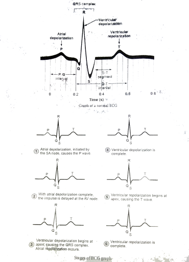
ST Segment : It lie between the QRS complex and T wave. It is the time during which all regions of the ventricles are completely depolarised and reflects the long plateau phase before repolarisation. In the heart muscle, the prolonged depolarisation is due to retardation of K+ ettlux and is responsible for the plateau. The ST segment lasts for 0.09 sec.
Twave (ventricular depolarisation): It represents ventricular depolarisation. The duration of the T wave is longer than QRS complex because repolarisation takes place simultancousty throughout the ventricular depolarisatiom.
P Wave (atrial depolarisation): It is a small upward wave and indicates the depolarisation of the atria. This is the time taken for the excitation to spread through atria from SA node. Contraction of both atria lasts for around 0.8-1.0 sec.
PQ Interval (AV node delay): It is the onset of P wave to the onset of QRS complex. This is from the start of depolarisation of the atria to the beginning of ventricular depolarisation. It is the time taken for the impulse to travel from the atria to the ventricles (0.12-0.21 sec). It is the measure of AV conduction time.
QRS Complex (ventricular depolarisation): No separate wave for atrial depolarisation in the ECG is visible. Atrial depolarisation occurs simultaneously with the ventricular depolarization. The normal QRS complex lasts for 0.06-0.09 sec. QRS complex is shorter than the P wave, because depolarisation spreads through the Purkinjie fibres. Prolonged QRS wave indicates delayed conduction through the ventricle, often caused due to ventricular hypertrophy or due to a block in the branches of the bundle of His
ST Segment : It lie between the QRS complex and T wave. It is the time during which all regions of the ventricles are completely depolarised and reflects the long plateau phase before repolarisation. In the heart muscle, the prolonged depolarisation is due to retardation of K+ ettlux and is responsible for the plateau. The ST segment lasts for 0.09 sec.
Twave (ventricular depolarisation): It represents ventricular depolarisation. The duration of the T wave is longer than QRS complex because repolarisation takes place simultancousty throughout the ventricular depolarisatiom.

One destination to cover all your homework and assignment needs
Learn Practice Revision Succeed

Instant 1:1 help, 24x7
60, 000+ Expert tutors

Textbook solutions
Big idea maths, McGraw-Hill Education etc

Essay review
Get expert feedback on your essay

Schedule classes
High dosage tutoring from Dedicated 3 experts
Practice more questions on Transport in mammals
Question 1
Easy
Views: 5,138
Question 2
Medium
Views: 5,252
Question 4
Easy
Views: 5,605
Students who ask this question also asked
Question 1
Views: 5,807
Question 3
Views: 5,856
Question 4
Views: 5,507


Stuck on the question or explanation?
Connect with our Biology tutors online and get step by step solution of this question.
231 students are taking LIVE classes
| Question Text | Explain the recording of electrocardiogram.
|
| Topic | Transport in mammals |
| Subject | Biology |
| Class | Class 12 |
| Answer Type | Text solution:1 |
